The gallbladder inflammation, known medically as cholecystitis, is brought on when bile cannot flow out of the gallbladder because of a gallstone. The illness causes excruciating discomfort. Surgery is sometimes the best option for long-term symptom alleviation for chronic cases with severe problems. It's good news that you can function normally without a gallbladder.
Precisely What Is Cholecystitis?

The gallbladder, an organ in the form of a pear, is located under the liver in the right upper quadrant of your belly. Gallbladders are organs responsible for storing and releasing bile, a fluid produced by the liver that aids in fat digestion. Cholecystitis occurs when bacteria infect bile that has been stuck in the gallbladder. When gallstones prevent bile from leaving the gallbladder, bile builds up and becomes toxic.
How Can Gallstones Obstruct Bile Flow?
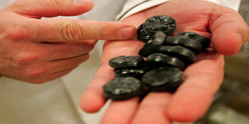
Gallstones are composed of hardened deposits of digestive fluids and can be as tiny as a grain of sand or as large as a golf ball. Cholesterol or pigment stones make up their composition. Cholesterol gallstones are often a more frequent yellowish green in appearance.
The liver produces bilirubin when it breaks down red blood cells, which is the primary component of pigment stones. Even if you have gallstones, it doesn't mean anything's wrong. If your gallstones are content to remain quietly in your gallbladder, causing you no discomfort, you don't need to have them removed.
In contrast, gallstones that pass through the gallbladder but become lodged in the duct system are a different story.
The Gallbladder: How Does It Function?
A network of tubes like the trunk and branches of a tree connects the gallbladder to the liver. The vents, or "branches," inside your liver are many. These twigs join the right and left hepatic ducts, the liver's two major arteries.
The common hepatic duct is the result of the union of these two ducts. The cystic duct is one major "tree limb" branching from the common hepatic duct. It has an open passageway that goes straight to the gallbladder.
Although the "tree trunk," or common hepatic duct, still exists, it is now known as the common bile duct. The duodenum of your small intestine is where your common bile duct ends.
How Can One Get Cholecystitis?
Gallstones are a common cause of cholecystitis because they obstruct the cystic duct and prevent bile from draining from the gallbladder. When this happens, your gallbladder swells and is at risk of bacterial infection.
Scarring of the bile ducts, decreased blood supply to the gallbladder, tumours that impede the outflow of bile, and viral infections that inflame the gallbladder are all less common causes.
How Can One Recognize Cholecystitis?
The symptoms may be sudden or ongoing.
Acute cholecystitis
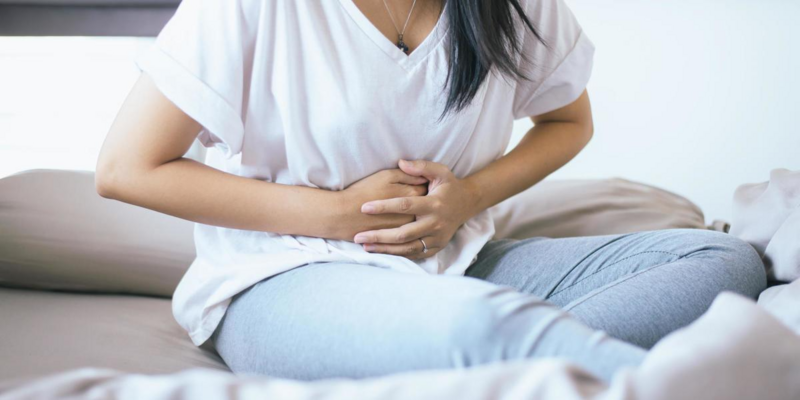
It occurs all of a sudden and continues to be quite painful. Gallstones are present in more than 95% of patients diagnosed with acute cholecystitis. When you first feel it, it's probably in your upper right abdomen, but it can go to your right shoulder blade and even your back. The worst pain hits about 15 minutes after eating and lasts for another 20. You must see a doctor about your persistent pain.
Chronic Cholecystitis
It signifies that you've been experiencing recurrent bouts of inflammation and discomfort. Compared to acute cholecystitis, the symptoms of chronic cholecystitis are often less severe and last for a shorter duration. Gallstones temporarily obstructing the cystic duct are the typical cause of the attacks' recurrence.
What Methods Are Used To Identify Cholecystitis?
Your doctor will inquire as to what exactly is wrong. Your white blood cell count and liver function may be among the tests they order if they suspect an infection. An elevated white blood cell count suggests the presence of infection, inflammation, or an abscess. Here are some examples of imaging procedures that could be prescribed:
Abdominal Ultrasound:
This diagnostic procedure analyses the gallbladder and bile ducts through ultrasonic waves. It aids in diagnosing gallbladder problems such as inflammation, gallstones, and gallbladder wall thickening or oedema.
Nuclear Hepatobiliary Imaging:
The radioactive material is administered for imaging purposes. A gamma camera tracks the radiation as it travels through the digestive system. If the doctor cannot inject the drug into the gallbladder, it is likely due to obstruction caused by cholecystitis.
MR Cholangiopancreatography:
The liver, gallbladder, and bile ducts, as well as pancreatic structures and chimneys, are all visible on this sort of MRI scan. Inflammation of the pancreas, gallstones, bile duct and gallbladder issues can all be shown.
How Does Gallbladder Surgery Recovery Go?
When the gallbladder is removed laparoscopically, the recovery process is often straightforward. Minor discomfort at the incision sites is expected following any surgical procedure. Most patients can go home soon after surgery without needing follow-up care. Recovery time and hospital days might increase if open surgery is performed.